UncategoriesLeg Bone Diagram / Luke Shaw expected to be out for up to nine months, expert ... : Joints of hand anterior view, lateral view, right hand.
Leg Bone Diagram / Luke Shaw expected to be out for up to nine months, expert ... : Joints of hand anterior view, lateral view, right hand.
Leg Bone Diagram / Luke Shaw expected to be out for up to nine months, expert ... : Joints of hand anterior view, lateral view, right hand.. Click now to learn more about the bones leg and knee anatomy: Visit kenhub for more skeletal system quizzes. For diagram showing its location relative to the fibula, tibia, patella, and other bones of the leg. Download the free graphic resources in the form of png, eps. Pngtree offers bone diagram png and vector images, as well as transparant background bone diagram clipart images and psd files.
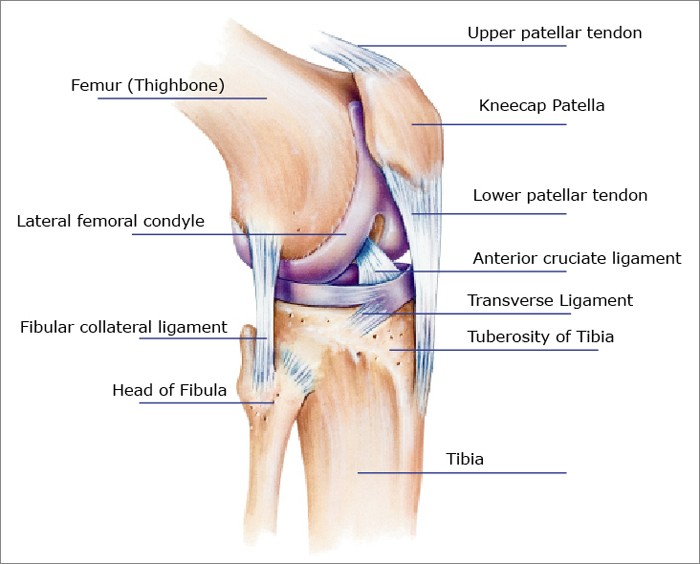
The bones of the leg are the femur, tibia, fibula and patella. While their parts are similar in general, their structure has been adapted to differing functions. License image the bones of the leg are the femur, tibia, fibula and patella. For diagram showing its location relative to the fibula, tibia, patella, and other bones of the leg. Learn how to draw the femur, patella, tibia, and fibula in this lesson!
Knee Pain: Symptoms, Causes, Treatments for Relief or ... from www.healthpages.org The bones of the leg are the femur, tibia, fibula and patella. Related online courses on physioplus. The foot bones shown in this diagram are the talus, navicular, cuneiform, cuboid. The humerus and the femur are corresponding bones of the arms and legs, respectively. When you stand or walk, all the weight of your upper body rests on them. The axial skeleton and the appendicular formed by the left and right hip bones, the pelvic girdle connects the lower limb (leg) bones to the axial. Visit kenhub for more skeletal system quizzes. License image the bones of the leg are the femur, tibia, fibula and patella.
Health diagram bone skeleton leg knee science anchor chart human human body.
He leg's main function in the human is for use the leg bones diagrams to learn the names of the leg bones and leg anatomy. The foot bones shown in this diagram are the talus, navicular, cuneiform, cuboid. Your leg bones are the longest and strongest bones in your body. Blood vessels and nerves enter the bone. Joints of hand anterior view, lateral view, right hand. Normal leg bones are relatively straight, but those affected by paget's disease are porous and figure 9. Cheek bone (zygoma) upper jaw (maxilla). Bone diagram barca fontanacountryinn com. Each leg is made up of four bones. Distal end of right humerus. Joints of hand anterior view, lateral view, right hand. The femur, or thighbone, is the longest and largest bone in the human body. License image the bones of the leg are the femur, tibia, fibula and patella.
Normal leg bones are relatively straight, but those affected by paget's disease are porous and figure 9. The foot bones shown in this diagram are the talus, navicular, cuneiform, cuboid, metatarsals and calcaneus. Leg femur diagram data wiring diagram today. Cheek bone (zygoma) upper jaw (maxilla). Learn vocabulary, terms and more with flashcards, games and other study tools.
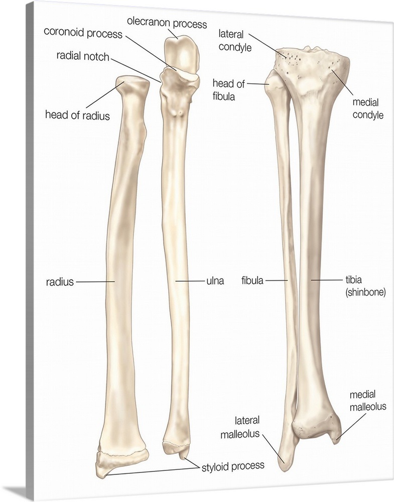
Comparison of bones of forearm and lower leg - anterior ... from static.greatbigcanvas.com Learn how to draw the femur, patella, tibia, and fibula in this lesson! Master leg and knee anatomy using our topic page. The bones of the leg are the femur, tibia, fibula and patella. Lower jaw (mandible) collar bone. Want to learn more about it? Your leg bones are the longest and strongest bones in your body. Pngtree offers bone diagram png and vector images, as well as transparant background bone diagram clipart images and psd files. Click now to learn more about the bones leg and knee anatomy:
Related online courses on physioplus.
The axial skeleton and the appendicular formed by the left and right hip bones, the pelvic girdle connects the lower limb (leg) bones to the axial. Normal leg bones are relatively straight, but those affected by. Learn how to draw the femur, patella, tibia, and fibula in this lesson! Quizzes on human skeletal system anatomy, bone anatomy, and bone markings. The femur, or thighbone, is the longest and largest bone in the human body. Your leg bones are the longest and strongest bones in your body. Bone diagram barca fontanacountryinn com. These bones have a marrow, but not a bone marrow cavity. New users enjoy 60% off. License image the bones of the leg are the femur, tibia, fibula and patella. While their parts are similar in general, their structure has been adapted to differing functions. Want to learn more about it? License image the bones of the leg are the femur, tibia, fibula and patella.
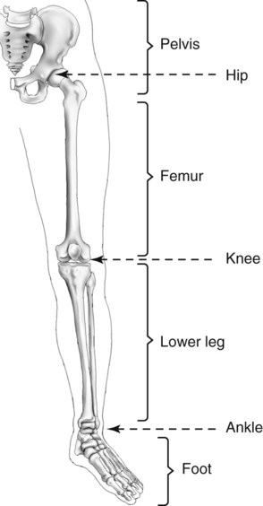
Visit kenhub for more skeletal system quizzes. The human leg, in the general word sense, is the entire lower limb of the human body, including the foot, thigh and even the hip or gluteal region. He leg's main function in the human is for use the leg bones diagrams to learn the names of the leg bones and leg anatomy. Time to jump right into the biggest and strongest bones in the human body. Diagram of blood and nerve supply to bone.
Lower Limb and Pelvis | Radiology Key from radiologykey.com Learn how to draw the femur, patella, tibia, and fibula in this lesson! Joints of hand anterior view, lateral view, right hand. Disposition of rotator cuff muscles diagram. Download the free graphic resources in the form of png, eps. Related online courses on physioplus. Human legs, modeled at anatomynext.com, based on radiology scans, theime atlas of anatomy, and expert advise. New users enjoy 60% off. Blood vessels and nerves enter the bone.
Click now to learn more about the bones leg and knee anatomy:
The bones of the leg are the femur, tibia, fibula and patella. Joints of hand anterior view, lateral view, right hand. For diagram showing its location relative to the fibula, tibia, patella, and other bones of the leg. Related online courses on physioplus. Human bone diagram wiring diagrams click. Cheek bone (zygoma) upper jaw (maxilla). The foot bones shown in this diagram are the talus, navicular, cuneiform, cuboid. Learn how to draw the femur, patella, tibia, and fibula in this lesson! Distal end of right humerus. Your leg bones are the longest and strongest bones in your body. Each leg is made up of four bones. The foot bones shown in this diagram are the talus, navicular, cuneiform, cuboid. Download 2,751 bone diagram stock illustrations, vectors & clipart for free or amazingly low rates!